43 simple microscope diagram with labels
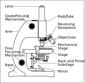
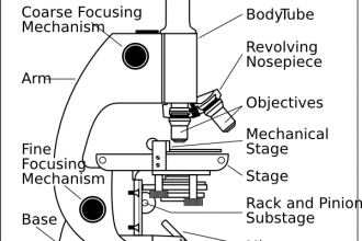
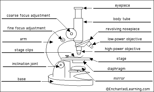
Labelled Diagram of Compound Microscope The below mentioned article provides a labelled diagram of compound microscope. Part # 1. The Stand: The stand is made up of a heavy foot which carries a curved inclinable limb or arm bearing the body tube. The foot is generally horse shoe-shaped structure (Fig. 2) which rests on table top or any other surface on which the microscope in kept. Simple Microscope Definition, Magnification, Parts And Uses - BYJUS Following are the parts of the simple microscope with their functions: Eyepiece: It is the lens that is used to study the samples and is placed at the top. It has a magnification of 10X to 15X. Base: This provides support to the microscope. Tube: This is used to connect the eyepiece to the objective lenses.
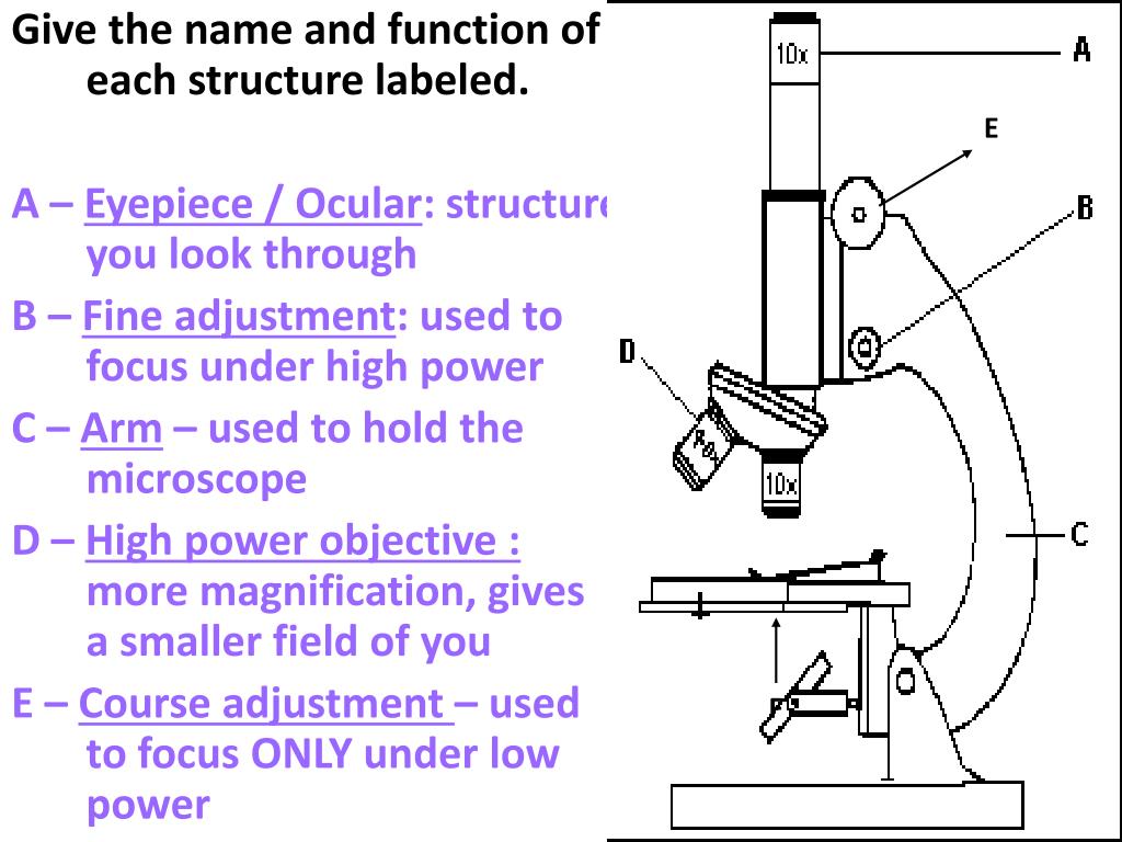
Microscope Labeling - The Biology Corner Microscope Labeling. Shannan Muskopf May 31, 2018. This simple worksheet pairs with a lesson on the light microscope, where beginning biology students learn the parts of the light microscope and the steps needed to focus a slide under high power. The labeling worksheet could be used as a quiz or as part of direct instruction where students label the microscope as you go over what each part is used for.
Simple microscope diagram with labels
Compound Microscope Parts - Labeled Diagram and their Functions There are two major optical lens parts of a microscope: Eyepiece (10x) and Objective lenses (4x, 10x, 40x, 100x). Total magnification power is calculated by multiplying the magnification of the eyepiece and objective lens. The illuminator provides a source of light. The light is focused by the condenser and passing through the specimen placed ... Parts of a microscope with functions and labeled diagram - Microbe Notes Head - This is also known as the body. It carries the optical parts in the upper part of the microscope. Base - It acts as microscopes support. It also carries microscopic illuminators. Arms - This is the part connecting the base and to the head and the eyepiece tube to the base of the microscope. Microscope Labeling - The Biology Corner Students label the parts of the microscope in this photo of a basic laboratory light microscope. Can be used for practice or as a quiz. ... The type of microscope used in most science classes is the _____ microscope. 18. You should carry the microscope by the _____ and the _____. 19. The objectives are attached to what part of the microscope ...
Simple microscope diagram with labels. Parts of a Simple Microscope - Labeled (with diagrams) image 5: A modern simple microscope with the different parts labeled. image source: laboratoryinfo.com The optical parts of a simple microscope are centered on the specimen - lighting, and magnification. Microscope Label Worksheets & Teaching Resources | TpT 48. $3.75. PDF (4.1 MB) This packet contains 13 community helper puzzles and labeling activities. These puzzles are a fun hands-on way to learn the names of some community helper parts/tools. Students can cut and glue the puzzle pieces onto the helper and use the labeling sheets to record each part. The packet also in. Labeling the Parts of the Microscope | Microscope World Resources Labeling the Parts of the Microscope. This activity has been designed for use in homes and schools. Each microscope layout (both blank and the version with answers) are available as PDF downloads. You can view a more in-depth review of each part of the microscope here. A Study of the Microscope and its Functions With a Labeled Diagram ... To better understand the structure and function of a microscope, we need to take a look at the labeled microscope diagrams of the compound and electron microscope. These diagrams clearly explain the functioning of the microscopes along with their respective parts. Man's curiosity has led to great inventions. The microscope is one of them.
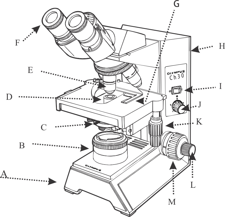
Microscope Types (with labeled diagrams) and Functions Simple microscope labeled diagram. Simple microscope functions. It is used in industrial applications like: Watchmakers to assemble watches; Cloth industry to count the number of threads or fibers in a cloth; Jewelers to examine the finer parts of jewelry; Miniature artists to examine and build their work; Also used to inspect finer details on products Compound Microscope Parts, Functions, and Labeled Diagram Compound Microscope Definitions for Labels. Eyepiece (ocular lens) with or without Pointer: The part that is looked through at the top of the compound microscope. Eyepieces typically have a magnification between 5x & 30x. Monocular or Binocular Head: Structural support that holds & connects the eyepieces to the objective lenses. Simple Microscope: Definition, Principle, Parts, And Uses A simple microscope is a rudimentary magnification device that is capable of visibly enlarging small objects, so they can be viewed and studied in better detail. It was invented in the late 16th century, and is still being widely used today. Simple microscopes have a wide range of applications in various fields. 16 Parts of a Compound Microscope: Diagrams and Video Once you have an understanding of the parts of the microscope it will be much easier to navigate around and begin observing your specimen, which is the fun part! The 16 core parts of a compound microscope are: Head (Body) Arm. Base. Eyepiece. Eyepiece tube.
Simple Microscope- Definition, Principle, Magnification, Parts ... A simple microscope is one that uses a single lens for magnification, such as a magnifying glass while a compound microscope uses several lenses to enhance the magnification of an object. It uses a lens to enlarge an object through angular magnification alone, giving the viewer an erect enlarged virtual image. The use of a single convex lens or ... Parts of the Microscope with Labeling (also Free Printouts) 5. Knobs (fine and coarse) By adjusting the knob, you can adjust the focus of the microscope. The majority of the microscope models today have the knobs mounted on the same part of the device. Image 5: The circled parts of the microscope are the fine and coarse adjustment knobs. Picture Source: bp.blogspot.com. Microscope Parts and Functions Microscope Parts and Functions With Labeled Diagram and Functions How does a Compound Microscope Work?. Before exploring microscope parts and functions, you should probably understand that the compound light microscope is more complicated than just a microscope with more than one lens.. First, the purpose of a microscope is to magnify a small object or to magnify the fine details of a larger ... Simple Squamous Epithelium under a Microscope with a Labeled Diagram ... Simple columnar epithelium labeled. This is a labeled diagram of a simple columnar epithelium under a light microscope. I tried to show you both ciliated and nonciliated simple columnar epithelium. These diagrams show the cilia on the cell surface, rectangular cell, and elongated nucleus.
Microscope With Labels clip art (111146) Free SVG Download / 4 Vector
Label the Microscope Diagram | Download Scientific Diagram - ResearchGate Gram staining was performed using compound microscope according to the procedure described by Petersen et al., 2016 [8]. The gram positive and gram negative bacteria were identified based on ...

Simple Cuboidal
Simple Microscope - Definition, Types, Working Principle & Formula 1. Simple microscope comprises a biconvex lens used as a magnifying glass. Compound microscope comprises 2 or more convex lenses where one lens is the eyepiece and the other one is the objective lens. 2. Natural light is the source to see the object. An illuminator is a source to see the object. 3.
Microscope Diagram With Labels And Functions - Micropedia
Simple Microscope - Parts, Functions, Diagram and Labelling Parts of the optical parts are as follows: Mirror - A simple microscope has a plano-convex mirror and its primary function is to focus the surrounding light on the object being examined. Lens - The biconvex lens is placed above the stage and its function is to magnify the size of the object being examined.
Foundations - Histology Epithelia and Skin - Embryology
Microscope Drawing Easy with Label - YouTube In this video I go over a microscope drawing that is easy with label. There is a blank copy at the end of the video to review on your own. A great way to s...
Get Practice Labeling Parts Of A Microscope Gif - DirectScot
Microscope With Labels clip art | Microscope parts, Scientific method ... Labeled microscope diagram Biological Science Picture Directory - Pulpbits.net. Print a microscope diagram, microscope worksheet, or practice microscope quiz in order to learn all the parts of a microscope. These parts of a microscope printables include word searches, crossword puzzles, and vocabulary worksheets.

11 Best Images of Cell Labeling Worksheet Answers - Cell Cycle and Mitosis Worksheet Answers ...
Microscope, Microscope Parts, Labeled Diagram, and Functions • Step 1: Connect the light microscope to a power source in step one. You can skip this step if your microscope has a mirror instead of an illuminator. Instead, look for a location with plenty of natural light. • Step 2: Rotate the revolving nosepiece so that the lowest objective lens is in place. • Step 3: Install your specimen on the stage. But first, make sure your specimen is adequately protected by placing a coverslip on top of it.
1.1 Labelling Microscope - Labelled diagram
Parts of a Microscope Labeling Activity - Storyboard That Knowing the names of the different parts of the microscope is essential to be able to use one properly. Create a poster that labels the parts of a microscope and includes descriptions of what each part does. Click "Start Assignment". Use a landscape poster layout (large or small). Search for a diagram of a microscope.

Microscope Unlabelled Diagram - Micropedia
Simple Microscope - Diagram (Parts labelled), Principle, Formula and Uses Simple microscope is a magnification apparatus that uses a combination of double convex lens to form an enlarged, erect image of a specimen. The working principle of a simple microscope is that when a lens is held close to the eye, a virtual, magnified and erect image of a specimen is formed at the least possible distance from which a human eye ...
Dissecting Microscope Labeled Diagram - Micropedia
Label the microscope — Science Learning Hub Label the microscope. Use this interactive to identify and label the main parts of a ...

The Microscope: Create a Labelled Diagram | Teaching Resources
Microscope labeled diagram - SlideShare Microscope labeled diagram 1. The Microscope Image courtesy of: Microscopehelp.com Basic rules to using the microscope 1. You should always carry a microscope with two hands, one on the arm and the other under the base. 2. You should always start on the lowest power objective lens and should always leave the microscope on the low power lens ...
Plant cell Structure: Plant cell parts, Organelles and their functions and Diagram
Microscope Poster - Diagram with Labels | Teach Starter A poster containing a diagram with labels showing the key parts of a microscope. In Science it is important that students know how to use a variety of tools when conducting scientific experiments and inquiry. This poster focuses on the microscope and highlights its key parts. There are two print options available for this poster: Print on tabloid paper to display around your school's science lab or in the experiment areas of your classroom.
Microscope labelling 11 - Teaching resources
Microscope Labeling - The Biology Corner Students label the parts of the microscope in this photo of a basic laboratory light microscope. Can be used for practice or as a quiz. ... The type of microscope used in most science classes is the _____ microscope. 18. You should carry the microscope by the _____ and the _____. 19. The objectives are attached to what part of the microscope ...

Clipart Panda - Free Clipart Images
Parts of a microscope with functions and labeled diagram - Microbe Notes Head - This is also known as the body. It carries the optical parts in the upper part of the microscope. Base - It acts as microscopes support. It also carries microscopic illuminators. Arms - This is the part connecting the base and to the head and the eyepiece tube to the base of the microscope.

29 best Anatomy and Physiology images on Pinterest | Physiology, Anatomy and Anatomy reference
Compound Microscope Parts - Labeled Diagram and their Functions There are two major optical lens parts of a microscope: Eyepiece (10x) and Objective lenses (4x, 10x, 40x, 100x). Total magnification power is calculated by multiplying the magnification of the eyepiece and objective lens. The illuminator provides a source of light. The light is focused by the condenser and passing through the specimen placed ...
Biology 521 Resources

Pin on Diagrams/Charts/Maps

Labelled Diagram Of A Tick - Top Label Maker

Cell organelles | Cells: the basic units of life | Siyavula
Post a Comment for "43 simple microscope diagram with labels"