40 structure of the heart with labels
Structure of the Heart | SEER Training The human heart is a four-chambered muscular organ, shaped and sized roughly like a man's closed fist with two-thirds of the mass to the left of midline. The heart is enclosed in a pericardial sac that is lined with the parietal layers of a serous membrane. The visceral layer of the serous membrane forms the epicardium. Layers of the Heart Wall Label the Heart - The Biology Corner A simple heart diagram with arrows and boxes for students to practice labeling the chambers and major vessels. Name:_____Date: _____ Label the Heart. Word Bank: Left Atrium | Right Atrium | Left Ventricle | Right Ventricle Aorta | Pulmonary Veins | Pulmonary Artery | Superior Vena Cava | Inferior Vena Cava Bicuspid Valve | Tricuspid Valve ...
Diagrams, quizzes and worksheets of the heart | Kenhub Labeled heart diagrams Take a look at our labeled heart diagrams (see below) to get an overview of all of the parts of the heart. Once you're feeling confident, you can test yourself using the unlabeled diagrams of the parts of the heart below. Labeled heart diagram showing the heart from anterior Unlabeled heart diagrams (free download!)

Structure of the heart with labels
Label the heart — Science Learning Hub Label the heart Interactive Add to collection In this interactive, you can label parts of the human heart. Drag and drop the text labels onto the boxes next to the diagram. Selecting or hovering over a box will highlight each area in the diagram. Right ventricle Right atrium Left atrium Pulmonary artery Left ventricle Pulmonary vein Semilunar valve Heart Diagram with Labels and Detailed Explanation - BYJUS Diagram of Heart. The human heart is the most crucial organ of the human body. It pumps blood from the heart to different parts of the body and back to the heart. The most common heart attack symptoms or warning signs are chest pain, breathlessness, nausea, sweating etc. The diagram of heart is beneficial for Class 10 and 12 and is frequently ... Structure of the Heart | The Franklin Institute Structure of the Heart Although most people know that the human heart doesn't bear much resemblance to a heart drawn on a Valentine's Day card, the image can still be a useful way to learn and remember the parts of the heart. The heart consists of four chambers: two atria on the top and two ventricles on the bottom.
Structure of the heart with labels. Label the Heart Diagram | Quizlet Label the Heart STUDY Learn Write Test PLAY Match Created by bluesas9 Terms in this set (15) Superior Vena Cava ... Right Ventricle ... Left Atrium ... Atrioventricular/Tricuspid Valve ... Atrioventricular/Mitral Valve ... Septum ... Right Atrium ... Semi-lunar Valves ... Left Pulmonary Veins ... Right Pulmonary Veins ... Left Pulmonary Arteries Structure Of The Heart | A-Level Biology Revision Notes The heart is a hollow muscular organ that lies in the middle of the chest cavity. It is enclosed in the pericardium, which protects the heart and facilitates its pumping action. The heart is divided into four chambers: The two atria (auricles): these are the upper two chambers. They have thin walls which receive blood from veins. The Heart - Science Quiz - GeoGuessr The Heart - Science Quiz Home >> Seterra Anatomy and Science Quizzes >> The Heart The Heart - Science Quiz Aorta, Aortic valve, Left atrium, Left ventricle, Mitral valve, Pulmonary artery, Pulmonary valve, Pulmonary vein, Right atrium, Right ventricle, Septum, Superior vena cava, Tricuspid valve (13) Create custom quiz Labelling the heart — Science Learning Hub Blood transports oxygen and nutrients to the body. It is also involved in the removal of metabolic wastes. In this interactive, you can label parts of the human heart. Drag and drop the text labels onto the boxes next to the diagram. Selecting or hovering over a box will highlight each area in the diagram.
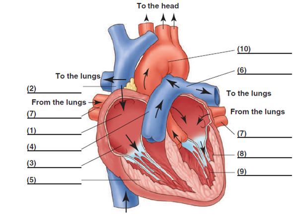
The Anatomy of the Heart - Quiz 1 - Free Anatomy Quiz The heart - an image of the heart with blank labels attached The circulatory system - upper body image, with blank labels attached The circulatory system - lower body image, with blank labels attached The circulatory system - a PDF file of the upper and lower body for printing out to use off-line Articles : How to Draw the Internal Structure of the Heart (with Pictures) To draw the internal structure of a human heart, follow the steps below. Part 1 Finding a Diagram 1 To find a good diagram, go to Google Images, and type in "The Internal Structure of the Human Heart". Find an image that displays the entire heart, and click on it to enlarge it. 2 Find a piece of paper and something to draw with. The Mammalian Heart | Biology for Majors II | | Course Hero The heart is composed of three layers; the epicardium, the myocardium, and the endocardium, illustrated in Figure 1. The inner wall of the heart has a lining called the endocardium.The myocardium consists of the heart muscle cells that make up the middle layer and the bulk of the heart wall. The outer layer of cells is called the epicardium, of which the second layer is a membranous layered ... Ch. 19 Circulatory System- heart Flashcards | Quizlet Place the labels in order denoting the flow of blood through the pulmonary circuit beginning with the right atrium and ending in the left atrioventricular valve. The first and last structures are given. Right atrium 1. tricuspid valve 2. right ventricle 3. pulmonary valve 4. pulmonary trunk 5. pulmonary artery 6. lungs 7. pulmonary vein
20 POINTS AVAILABLE Identify the structures of the heart. Label A Label ... Septum is the tissue that separates left and right ventricles while aorta is the main artery which carry blood from heart to tissues. Thus, label A is pulmonary valve, label B is tricuspid valve, label C is septum, label D is bicuspid valve, label E is aortic valve and label F is aorta. For more information about structure of heart, visit: Human Heart (Anatomy): Diagram, Function, Chambers, Location in Body The heart is a muscular organ about the size of a fist, located just behind and slightly left of the breastbone. The heart pumps blood through the network of arteries and veins called the... Heart Labeling Quiz: How Much You Know About Heart Labeling? Here is a Heart labeling quiz for you. The human heart is a vital organ for every human. The more healthy your heart is, the longer the chances you have of surviving, so you better take care of it. Take the following quiz to know how much you know about your heart. Questions and Answers 1. What is #1? 2. What is #2? 3. What is #3? 4. What is #4? Heart Diagram with Labels and Detailed Explanation There are four chambers of the heart. The upper two chambers are the auricles and the lower two are called ventricles. There are four main valves of the human heart- aortic valve, mitral valve, pulmonary valve and tricuspid valve. They help prevent backflow of the blood.
![Untitled Document [www.stritch.luc.edu]](http://www.stritch.luc.edu/lumen/MedEd/Radio/curriculum/structure/Plain_xray/normal_male_pa_labelled.jpg)
Untitled Document [www.stritch.luc.edu]
Human Heart - Anatomy, Functions and Facts about Heart Label the Heart Diagram below: Practice your understanding of the heart structure. Drag and drop the correct labels to the boxes with the matching, highlighted structures. Instructions to use: Hover the mouse over one of the empty boxes. One part in the image gets highlighted.
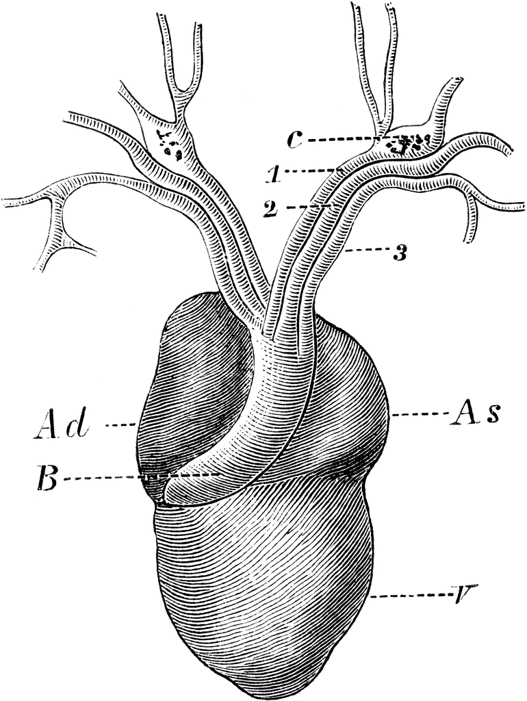
Heart of a Frog | ClipArt ETC
ERIC - ED202539 - Health Instruction Packages: Cardiac Anatomy., 1977 Text, illustrations, and exercises are utilized in these five learning modules to instruct nurses, students, and other health care professionals in cardiac anatomy and functions and in fundamental electrocardiographic techniques. The first module, "Cardiac Anatomy and Physiology: A Review" by Gwen Phillips, teaches the learner to draw and label the parts of the heart and its impulse conduction ...
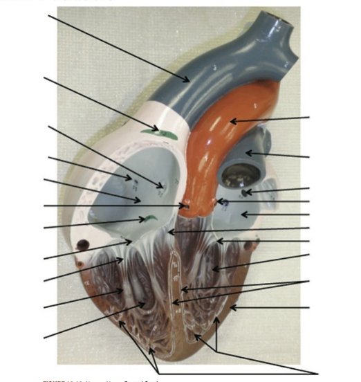
Solved: Label The Following Structures Of The Heart | Chegg.com
Layers of the heart: Epicardium, myocardium, endocardium - Kenhub The myocardium is functionally the main constituent of the heart and the thickest layer of all three heart layers. It is a muscle layer that enables heart contractions. Histologically, the myocardium is comprised of cardiomyocytes.Cardiomyocytes have a single nucleus in the center of the cell, which helps to distinguish them from skeletal muscle cells that have multiple nuclei dispersed in the ...

your lungs and how breathing works diagram of the human respiratory system infographic by ross ...
Anatomy of the heart and coronary arteries (coronary CT) - IMAIOS Anatomy of the human heart and coronaries: how to view anatomical structures. This tool provides access to an MDCT atlas in the 4 usual planes, allowing the user to interactively discover the heart anatomy. The images are labeled, providing an important medical and anatomical tool. The quiz mode makes it possible to evaluate the user's progress.

Eclectic Photography Project: Day 144 - strolling around Eureka, CA
Human Heart - Diagram and Anatomy of the Heart - Innerbody Because the heart points to the left, about 2/3 of the heart's mass is found on the left side of the body and the other 1/3 is on the right. Anatomy of the Heart Pericardium. The heart sits within a fluid-filled cavity called the pericardial cavity. The walls and lining of the pericardial cavity are a special membrane known as the pericardium.

The human egg cell explained for egg donors | Altrui
The Anatomy of the Heart, Its Structures, and Functions The heart is the organ that helps supply blood and oxygen to all parts of the body. It is divided by a partition (or septum) into two halves. The halves are, in turn, divided into four chambers. The heart is situated within the chest cavity and surrounded by a fluid-filled sac called the pericardium. This amazing muscle produces electrical ...

34 Label The Heart Diagram - Labels Database 2020
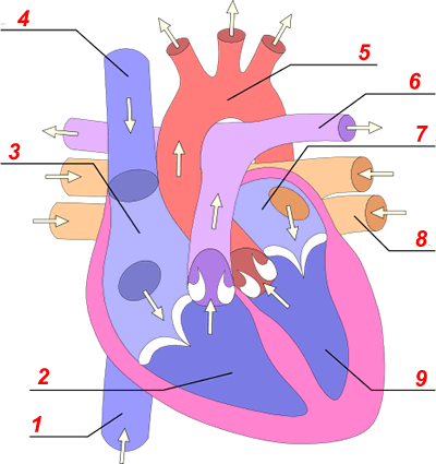
Label the Heart Shows a picture of a heart with letters and blanks for practice with labeling the parts of the heart and tracing the flow of blood within the heart.
Structure and Function of the Heart
Heart Anatomy Labeling Game - PurposeGames.com This is an online quiz called Heart Anatomy Labeling Game There is a printable worksheet available for download here so you can take the quiz with pen and paper. Your Skills & Rank Total Points 0 Get started! Today's Rank -- 0 Today 's Points One of us! Game Points 19 You need to get 100% to score the 19 points available Actions
Solved: Label the following structures of the heart by writing ... | Chegg.com
The structure of the heart - Structure and function of the heart ... It is located in the middle of the chest and slightly towards the left. The heart is a large muscular pump and is divided into two halves - the right-hand side and the left-hand side. The...

What are Arteries? - Function & Definition - Video & Lesson Transcript | Study.com
Heart Blood Flow | Simple Anatomy Diagram, Cardiac Circulation ... - EZmed Step 2 involves the left atrium, the chamber of the heart that receives oxygenated blood from the lungs via the pulmonary veins. 3. Mitral Valve Step 3 involves the mitral valve. During diastole, when the heart is relaxed and filling with blood, the oxygenated blood from the left atrium will flow to the left ventricle.
Free Heart Diagram Unlabeled, Download Free Heart Diagram Unlabeled png images, Free ClipArts on ...
147 Heart Anatomy With Labels Premium High Res Photos Browse 147 heart anatomy with labels stock photos and images available, or start a new search to explore more stock photos and images. heart blood flow - heart anatomy with labels stock illustrations. human heart anatomy. blood flow - heart anatomy with labels stock illustrations. circulatory system, diagram - heart anatomy with labels stock ...
Label Heart Structure
Heart: Anatomy and Function - Cleveland Clinic The parts of your heart are like the parts of a house. Your heart has: Walls. Chambers (rooms). Valves (doors). Blood vessels (plumbing). Electrical conduction system (electricity). Heart walls Your heart walls are the muscles that contract (squeeze) and relax to send blood throughout your body.
Post a Comment for "40 structure of the heart with labels"