38 sperm cell diagram with labels
Draw the diagram of human sperm and label its parts. ... Draw the diagram of human sperm and label its parts. Write few lines about it. Medium Solution Verified by Toppr The sperm cells are the haploid gametes which are produced in the male. There are different parts of the sperm cell. (a) Acrosome: This structure contains enzymes used for penetrating the female egg. Diagram and label sperm cell - Quizlet Only $2.99/month Diagram and label sperm cell STUDY Learn Flashcards Write Spell Test PLAY Match Gravity Created by Ike_SandersonTEACHER Terms in this set (4) Midsection of sperm contains mitochondria Sperm nucleus Contains haploid chromosomes Acrosome A vesicle at the tip of a sperm cell that helps the sperm penetrate the egg Flagellum
Cambridge Assessment International Education ... - Save My Exams 29 A human zygote is a diploid cell. Which statement about human diploid cells is correct? A They do not have a nucleus. B They fuse to form gametes. C The nucleus contains a single set of chromosomes. D The nucleus contains two sets of chromosomes. 30 Which feature allows the sperm to dissolve the jelly coating of the egg cell? A acrosome B ...

Sperm cell diagram with labels
Male Reproductive System: Labeled Diagram of Organs - Study.com The epididymis is a coiled tube present on each testicle that hosts sperm after they are produced. Sperm stored in the epididymis undergo further maturation, acquire motility, and reside there... chap 27 and 28 Flashcards | Quizlet Drag the labels onto the diagram to identify the stages of spermatogenesis. zygote The fertilized egg, or __________, appears as a single cell surrounded by a fertilization membrane and a jellylike membrane. Sperm Diagram Stock Photos, Pictures & Royalty-Free Images - iStock Human Sperm cell Anatomy Comparison between normal and low sperm count Female reproductive system, image diagram Penis or Male Reproductive System is a 3D illustration. Human fertility Embryo development concept. Insemination and fertalization. Female and male egg cell icon. Human sexual reproductive system and pregnancy flat vector illustration.
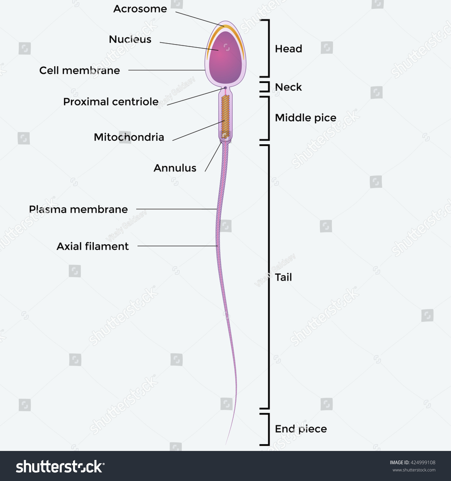
Sperm cell diagram with labels. Sperm Cells - Definition, Function, Structure, Adaptations ... The head of the sperm measures 2.5 to 3.5 um in diameter and 4.0 to 5.5 um in length (um=micrometers). This results in a 1.50 to 1.70 length to width ratio They have a well-developed acrosome that covers 40 to 70 percent of the oval shaped head A slim middle section (body) that is approximately the same length as the head Draw a Diagram of the Microscopic Structure of Human ... Answer in Brief Diagram Draw a diagram of the microscopic structure of human sperm. Label the following parts in it and write their functions (a) Acrosome (b) Nucleus (c) Middle piece Draw a diagram of a mature human sperm. Label any three parts and write their functions. Explain the structure of human sperm with labelled diagram. The Mitochondrion - Molecular Biology of the Cell - NCBI ... Thus, the mitochondria in some cells form long moving filaments or chains. In others they remain fixed in one position where they provide ATP directly to a site of unusually high ATP consumption—packed between adjacent myofibrils in a cardiac muscle cell, for example, or wrapped tightly around the flagellum in a sperm (Figure 14-6). Structure and parts of a sperm cell - inviTRA Structure and parts of a sperm cell 0 This labelled diagram shows the structure of a sperm cellin detail, which has the following parts: Head With its spheric shape, it consists of a large nucleus, which at the same time contains an acrosome. The nucleus contains the genetic information and 23 chromosomes.
Draw a labeled diagram of sperm. - SaralStudy Q:-With a neat diagram explain the 7-celled, 8-nucleate nature of the female gametophyte. Q:-What is oogenesis? Give a brief account of oogenesis. Q:-What is DNA fingerprinting? Mention its application. Q:-With a neat, labelled diagram, describe the parts of a typical angiosperm ovule. Q:-What is triple fusion? Where and how does it take place? Show well labeled diagram of the structure of human sperm and write ... Sperms are microscopic, motile cells that have a head, neck, middle section, and tail. (i) Head: It has a little acrosome in the front and a large nucleus in the back. The golgi body of the spermatid forms the acrosoma. (ii) Neck: Between the head and the middle section, there is a very short neck. […] Draw a diagram of the microscopic structure of human ... - Toppr The above diagram is of the sperm cell. (a) Acrosome: It contains enzymes used for penetrating the female egg. (b) Nucleus: Contains the genetic material that the sperm has to pass on, a haploid genome because it contains only one copy of each chromosome. Male reproductive: The Histology Guide - University of Leeds The production of sperm and eggs/ova (gametes) is a procedure called gametogenesis (spermatogenesis and oogenesis). Gametogenesis involves two rounds of meiotic cell division, in which one diploid cell gives rise to 4 haploid cells.. This diagram shows the processes involved in spermatogenesis. The germinal (seminiferous epithelium) of the seminiferous tubules contains spermatogenic cells and ...
Sperm - Wikipedia Sperm quantity and quality are the main parameters in semen quality, which is a measure of the ability of semen to accomplish fertilization.Thus, in humans, it is a measure of fertility in a man.The genetic quality of sperm, as well as its volume and motility, all typically decrease with age.. DNA damages present in sperm cells in the period after meiosis but before fertilization may be ... Draw the diagram of human sperm and label its parts ... - Toppr Draw the diagram of human sperm and label its parts. Write few lines about it. · The sperm cells are the haploid gametes which are produced in the male. There ... Sperm Cell, Egg Cell Diagram Label Worksheets (Differentiated) Three excellently differentiated worksheets. Engaging activity where pupils have to label the different parts of the male and femal gametes. Very well structured and scaffolded according to ability (from SEN to high ability). Excellent for visual learners. Compatible with all biology exam boards (including AQA, Edexcel, OCR). ch 8 mastering biology Flashcards | Quizlet Can you label the phases of the cell cycle? To review a crucial phase of the cell cycle, watch this BioFlix animation: Mitosis. Part A - The cell cycle Drag the pink labels onto the pink targets to identify the two main phases of the cell cycle. Then drag the blue labels onto the blue targets to identify the key stages that occur during those ...
Organelles of sperm
Year 7 - Science Revision Guide - Biology ... - Chauncy School Every month an ovum (egg cell) is released from an ovary into the oviduct. This is called OVULATION. If there are sperm cells in the oviduct the ovun may join with one of them. This is called FERTILISATION. The fertilised ovum then travels down to the uterus where it grows Into a baby. The diagram below shows what happens to the ovun after it is
Illustartion Showing Structure Sperm Cell Stock Vector 424999108 - Shutterstock
Sperm Cells Images | Free Vectors, Stock Photos & PSD Find & Download Free Graphic Resources for Sperm Cells. 500+ Vectors, Stock Photos & PSD files. Free for commercial use High Quality Images ... Diagram showing human sex cells on white background. brgfx. 8. Like. Collect. Save. In vitro fertilization flat elements. macrovector. 22. Like. Collect. Save. In vitro fertilization concept ...

Sperm Cells for Artificial Reproduction and Germ Cell Transplantation - European Urology Supplements
Specialised animal cells - Cell structure - BBC Bitesize The tail enables the sperm to swim. Sperm are the smallest cells in the body and millions of them are made. Egg cell. The cytoplasm contains nutrients for the growth of the early embryo. The ...

Reproduction flashcards | Quizlet
Sperm Diagram Stock Illustrations - 415 Sperm Diagram Stock ... Download 415 Sperm Diagram Stock Illustrations, Vectors & Clipart for FREE or amazingly low rates! ... Ocean depth zones infographic, vector illustration labeled diagram. Oceanography science educational graphic information. Depth at which sperm whales live and. ... Blue sperm cell vector illustration. 3d fertilisation isolated.
Structure of a sperm cell stock vector. Illustration of human - 43980330
Spermatogenesis Diagram & Function | What is the Process of Sperm ... Beneath the Sertoli cells are the spermatogonia, which are germ cells that will go through mitosis and ultimately create sperm. In humans, each day, roughly 25 million spermatogonia divide, and...

What is the structure of a mature human sperm cell? - Lifeeasy Biology: Questions and Answers
Diagram showing a mature human spermatozoon with its ... Proteins localized in acrosome, head, midpiece, and tail are mainly involved in capacitation and acrosomal reaction, spermatozoa-oocyte interaction (zona ...

Sperm Cell Diagram Labeled - ClipArt Best
Labeled Sperm Cell Pictures, Images and Stock Photos Browse 15 labeled sperm cell stock photos and images available, or start a new search to explore more stock photos and images. Newest results Receptionist labeling sample in a laboratory Prostate labeled vector illustration. Educational male anatomy... Cell potency. From Totipotent to Pluripotent, Multipotent, and...

Second Questions
Sperm Cell Labeled Diagram Stock Vector (Royalty Free) 200461103 ... Frequently used Trendsetter We're seeing significant engagement with this asset. Item ID: 200461103 Sperm Cell Labeled Diagram Formats EPS 6733 × 3563 pixels • 22.4 × 11.9 in • DPI 300 • JPG Contributor j joshya Similar images See all Assets from the same collection See all Similar video clips

Line Diagram Of A Sperm - ClipArt Best
Sperm Cell - The Definitive Guide | Biology Dictionary A sperm cell or spermatozoon is a gamete (sex cell) produced in the male reproductive tract. It is a motile cell with a single aim - to fertilize a female egg. Each sperm cell contains the entire genome of the male that produces it. In combination with the female genome contained within the egg, a zygote is formed - a single totipotent stem ...

Sperm Cell Diagram Labeled - ClipArt Best
Fertilization Diagram Stock Illustrations - Dreamstime Part of a flower biological diagram, vector illustration drawing with educational scheme. Labeled plant cross section with ovary, pistil, sepal and stamen. ... Sperm Cell of Human Body Anatomical Diagram. With all parts including head middle piece and tail neck mitochondrion nucleus plasma membrane for anatomy biology.
Reproductive System Worksheet Answers - WikiEducator
Draw the diagram of the human sperm and label its parts class 12 ... It is a microscopic structure, motile in nature, and fertilizes the female gamete, the egg. In mammals, the motile sperm travels with the help of a fluid known as semen. Complete step by step answer: - The human sperm can be divided into the head, the neck, the middle piece, and the tail. - The entire body is enveloped by a plasma membrane.
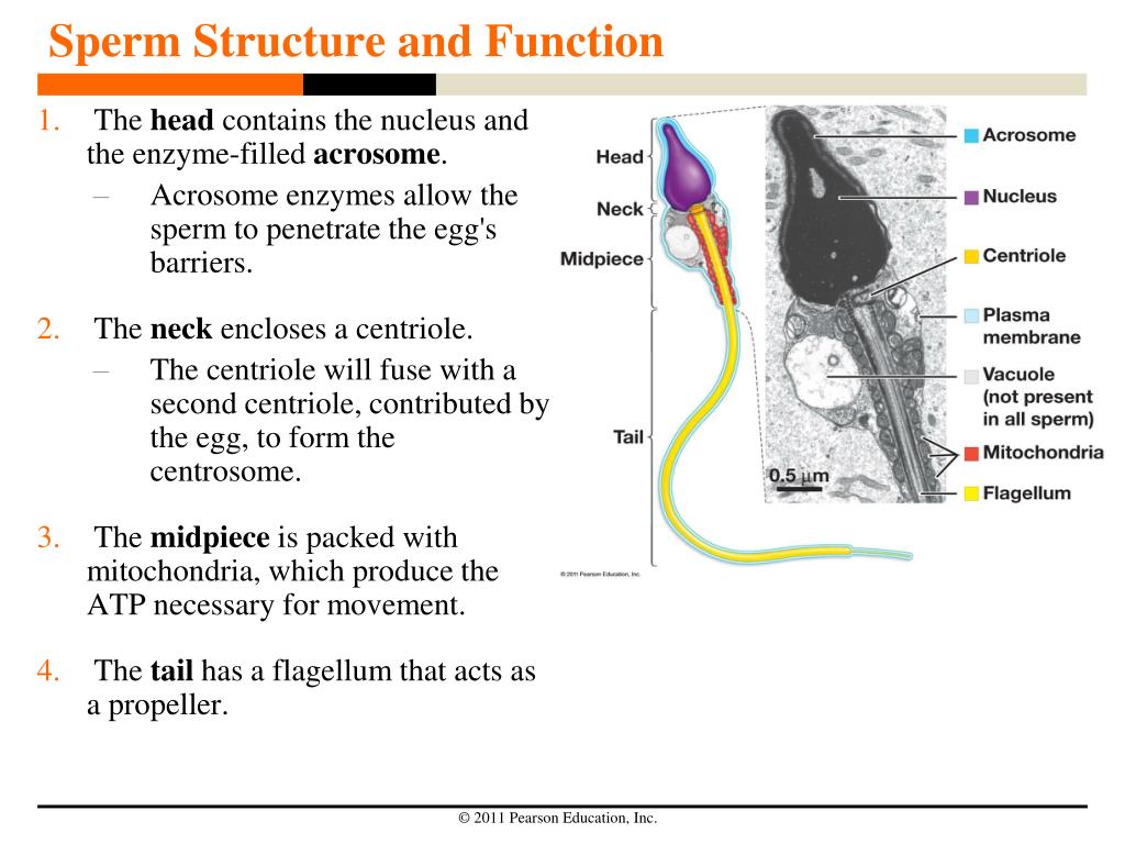
PPT - Sperm Structure and Function PowerPoint Presentation, free download - ID:4130804
Recurrent inversion polymorphisms in humans associate ... - Cell May 06, 2022 · Venn diagram depicts overlap by approach for 127 tested inversions. (C–E) Evidence for single (C, 17q21) and recurrent (D, 8p23.1 [distal part chr8:8225000-8301024]; E, 11p11) loci. Left: dendrograms (centroid hierarchical clustering method) show relationships among inverted and direct-oriented haplotypes.

Brenda's A & P Eportfolio: Objective 71 & 72: How sperm move and evolutionary fitness
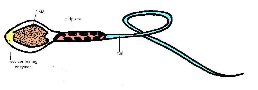
An overview of sperm anatomy | Legacy Sperm is the male sex cell, also known as a gamete. Measuring approximately 0.05 millimeter (0.002 inch) long, sperm cells are made up of a few distinct parts: the tail, made up of protein fibers, which helps it "swim" toward the egg the midpiece, or body, which contains mitochondria to power the sperm's movement

BIOLOGY CST PracticeReleased California State BiologyTest Questions. © California Department of ...
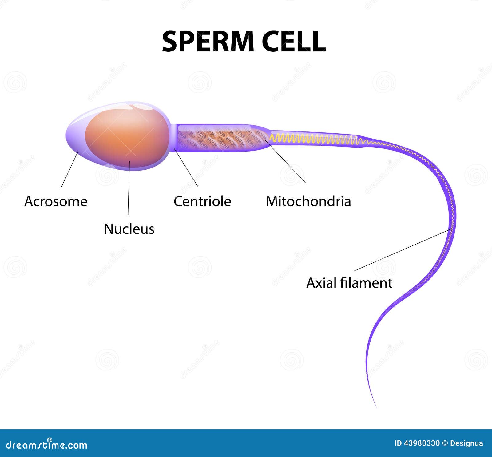
Draw the diagram of human sperm and label its parts ... - Toppr The sperm cells are the haploid gametes which are produced in the male. There are different parts of the sperm cell. (a) Acrosome: This structure contains ...
Post a Comment for "38 sperm cell diagram with labels"